Monday, January 31, 2011

Serendipity

Serendipity, fortunate discoveries by accident.

 
Serendipity is making fortunate discoveries by accident. Especially when one is looking for something unrelated, these lucky events can occur. In our daily life, sometimes we come across fortunate findings that makes us wonder. Usually, we phrase this as coincidence. Also in Physics and Science, in particular in fields as pharmacology and chemistry, there have been quite a few serendipitous discoveries. A famous example is Alexander Flemings discovery of penicillin. 


A creative serendip link

Links related to serendipity:
Rodin's The Thinker & 20 microns lasered sculpture

(I'm thinking about spending a blogpost about thinking)

Serendipity, fortunate findings makes us wonder.

Serendipity, mistakes are the portals of discoveries

Famous serendipitous examples:
  • The organic chemist Kekulé had a vision of a snake forming a circle, which led to his solution of closed chemical structures such as the cyclic compound benzene.
  • While the chemist Louis Pasteur was investigating the properties of sodium ammonium tartrate, he managed to separate the two optical isomers of the salt. His luck was twofold: it turned out to be the only racemate salt with this property and the room temperature at that day was just below the seperation point.
  • A chemist unintentionally, absorbed a small amount of LSD upon investigating it's properties, and had the first trip in history.
  • The observation of an apple falling from a tree, by Isaac Newton, was at the basis for a theory about the nature of gravitation. 
  • The discovery of X rays by Wilhelm Roentgen as radiation that could pass a lighttight cover.
  • Astronomers discovered the rings of Uranus from an observation of periodic brief disappearance of view.
  • During the repair of the Star Scan Machine, there was time to inspect photographic plate with the same "defects", a bulge in the planet's image which was actually the moon Charon of Pluto.
  • While searching for a nonconducting material, superconductivity was found  instead.
  • Inkjet printers were invented by accidently putting a hot iron on a pen, the ink was ejected from the pen's point a few moments later.
  • The microwave oven was invented after a peanut candy bar in a men's pocket was melted after exposure with radar waves.
  • Columbus was looking for a new way to India and landed in the America's.

Serendipitous example in our laboratory:
closed and open conformation of the PITP protein
For quite a number of trials we failed in improving the X-ray diffraction quality for crystals from a phospholipid-binding transfer protein in order to solve its molecular structure. Finally, we came up with the idea to exchange the bound lipid with a modified lipid having bromine as heavy atom to accomplish phasing. Phasing is essential to solve the crystal structure of a protein with low sequence homology. In the meantime the competitors scooped us by succeeding in solving the structure of the natural phospholipid containing protein (closed holo-form). Luckily, in a later stage, we revealed that the protein was not able to accomodate our modified phospholipid at all, probably due to steric hindrance. Nevertheless, we succeeded in elucidating the crystal structure of this phospholipid free protein (open apo-form), that turned out to be a representative model for the interesting membrane-bound conformation of the protein (see apo-PITP). In view of understanding the role of the  phospholipid binding protein, the model for phospholipid binding and membrane association had major implications for its function: from closed for transport to open for exchange. An example of fortunate discovery by accident.

Cybernetic serendipity, an exhibition of computer art
Serendipity, the art of making happy discoveries

Thursday, January 13, 2011

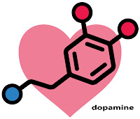
Dopamine

NL Dopamine.

Waarom een blogpost over dopamine? 
De laatste tijd wordt er steeds meer bericht over experimenten waarbij dopamine een belangrijke rol lijkt te spelen. Zo blijkt luisteren naar muziek de aanmaak van deze genotstof in het brein te kunnen stimuleren. Een ander onderzoek toonde aan dat als er 'Liefde' in het spel is, er veel aktiviteit aanwezig is in de zogenaamde beloningsgebieden van het brein waarin dopamine wordt afgegeven. Hierbij kunnen er delen van de hersenen uitgeschakeld raken of kan zelfs een gedeelte van het brein worden geblokkeerd. Dopamine speelt dus een grote rol bij het ervaren van genot, blijdschap en welzijn. De beloningsgebieden zijn blijkbaar gevoelig voor de aanwezige hoeveelheid van de genotstof dopamine. Helaas blijkt een onbalans in de aanmaak van dopamine een nadelige rol te spelen bij ADHD, schizofronie en de ziekte van Parkinson. Het leek me daarom de moeite waard om er iets in deze blogpost over te vermelden.

Volgens Wikipedia is dopamine is een verbinding die fungeert als neurotransmitter (en soms als hormoon) op diverse plaatsen in het lichaam. Het komt in het menselijk en dierlijk organisme ook voor als precursor van de hormonen adrenaline en noradrenaline. Het speelt een grote rol bij het ervaren van genot, blijdschap en welzijn. Positief affect, zoals een prettig gevoel na beloning, blijkt een gunstige uitwerking te hebben op cognitieve prestaties. Volgens de theorie van Ashby speelt hierbij een circuit in de hersenen een rol waarbij dopamine wordt vrijgemaakt in de basale ganglia en vervolgens getransporteerd naar de frontale hersengebieden. Mensen met de ziekte van Parkinson hebben een tekort aan dopamine. Ook bij ADHD is er waarschijnlijk sprake van een onbalans van dopamine. Dopamine speelt tenslotte ook een rol bij schizofrenie.

Muziek zorgt voor aanmaak genotstof in brein (NU.nl en Columnary).
Mensen kunnen intens genieten van het luisteren naar muziek, omdat er onder invloed van muziekklanken dopamine wordt aangemaakt in het menselijk brein.
 

Als mensen zich laten meevoeren door een mooi stuk muziek, of genieten van een specifiek muzikaal moment, worden die gevoelens zeer waarschijnlijk veroorzaakt door dopamine. Deze ‘genotstof’ die ook vrij komt onder invloed van seks, drugs en cafeïne, wordt vrijwel direct aangemaakt in het brein bij het luisteren naar muzikale klanken.
Kippenvel: Tijdens een onderzoek, waarbij hersenscans werden gemaakt van mensen die naar muziek luisterden bleek dat er tijdens deze momenten buitengewoon veel dopamine werd geproduceerd in de 'beloningscentra' van hun hersenen.
Hoogtepunten: De aanmaak van de genotstof nam al toe in de vijftien seconden die vooraf gingen aan de muzikale hoogtepunten. Maar ook als de proefpersonen luisterden naar muziek die niet onmiddellijk voor een kippenvelmoment zorgde, kwam er dopamine los in hun brein. De resultaten van het onderzoek suggereren dat ook mensen die wel genieten van muziek maar geen kippenvel krijgen, de effecten van dopamine ervaren.       
Instrumentele muziek: Bij de studie werd alleen gebruik gemaakt van instrumentale muziek. Daarmee is volgens de wetenschappers aangetoond dat de aanwezigheid van zang en songteksten niet noodzakelijk is om de productie van dopamine te stimuleren.
 
Liefde blokkeert gedeelte van brein (NU.nl).
Als verliefde mensen naar hun partner kijken, worden er delen van hun hersenen uitgeschakeld.

Er is gebleken dat liefde inderdaad echt 'blind' maakt. Passionele romantische liefde wordt meestal geactiveerd door visuele input en zorgt voor een allesoverheersende en gedesoriënteerde staat. Verder bleek dat er een specifiek patroon van hersengebieden actief werd als de proefpersonen keken naar hun geliefde. Vooral de zogenaamde beloningsgebieden waarin dopamine wordt afgegeven, vertoonden veel activiteit. 

Uit een geheel ander onderzoek naar het effect van vrouwentranen op mannen blijkt dat niet dopamine maar feromonen (chemische stoffen die boodschappen overbrengen tussen individuen van eenzelfde soort) betrokken zijn.

Extraverte mensen zijn creatiever (NU.nl).
Een extraverte persoonlijkheid is een voorwaarde voor het presteren bij het uitvoeren van creatieve taken. Gemiddeld genomen wordt er meer van de ‘genotstof’ dopamine aangemaakt in de hersenen van niet-introverte personen. Als ze daarbij ook nog in een goede stemming of flow verkeren, bereikt de productie van dopamine en het creatieve denkvermogen een maximaal niveau.  

Links: 

Sunday, January 9, 2011

Autophagy

Introduction to Autophagy.


1. Autophagy is a lysosomal degradation pathway that is essential for survival, differentiation, development, and homeostasis. Autophagy principally serves an adaptive role to protect organisms against diverse pathologies, including infections, cancer, neurodegeneration, aging, and heart disease. However, in certain experimental disease settings, the self-cannibalistic or, paradoxically, even the prosurvival functions of autophagy may be deleterious.
[4. Beth Levine et al., Autophagy in the Pathogenesis of Disease, Cell, 132, Issue 1, 27-42, 2008]

The Autophagic Pathway

2. Autophagy (Beth Levine: HHMI) is an evolutionarily conserved pathway that involves the lysosomal degradation of long-lived cellular proteins and organelles. Our laboratory identified the first mammalian autophagy protein, Beclin 1, in a screen for novel proteins that interact with the apoptosis inhibitor, Bcl-2. Work by our laboratory and others has established that autophagy participates in a number of fundamental biological processes and diseases. Using genetic approaches in different model organisms, we have shown that beclin 1 and autophagy function in tumor suppression, in development and lifespan extension, and in innate antiviral immunity. We have also found that the Bcl-2/Beclin 1 complex provides rheostatic control on autophagy and cell death. Our ongoing research aims to further identify the precise role of autophagy in antimicrobial host defense, in cancer biology, in cell death regulation, and in the prevention of aging and neurodegenerative diseases. To accomplish this goal, we are using a combination of biochemistry, structural biology, molecular biology, and genetic approaches in yeast, C. elegans, and mammalian systems.


3. Autophagy is the major cellular pathway for the degradation of long-lived proteins and cytoplasmic organelles.  It involves the rearrangement of subcellular membranes to sequester cargo for delivery to the lysosome where the sequestered material is degraded and recycled.  For many decades, it has been known that autophagy occurs in a wide range of eukaryotic organisms and in multiple different cell types during starvation, cellular and tissue remodeling, and cell death. However, until recently, the biological functions of autophagy have been largely unknown. The identification of a set of evolutionarily conserved genes that are essential for autophagy has opened up new frontiers for understanding the role of autophagy in diverse biological processes.  

Links:

Saturday, January 8, 2011

Membrane proteins

Membrane proteins.

Cell membrane

A membrane protein is a protein molecule that is attached to, or associated with the membrane of a cell or an organelle. More than half of all proteins interact with
membranes. 

Illustration of a Eukaryotic cell membrane

Biological membranes consist of a phospholipid bilayer and a variety of proteins that accomplish vital biological functions. Structural proteins are attached to microfilaments in the cytoskeleton which ensures stability of the cell. Cell adhesion molecules allow cells to identify each other and interact. Such proteins are involved in immune response, for example. Membrane enzymes produce a variety of substances essential for cell function. Membrane receptor proteins serve as connection between the cell's internal and external environments. Finally, transport proteins play an important role in the maintenance of concentrations of ions. These transport proteins come in two forms: carrier proteins and channel proteins. Carrier proteins are involved in using the energy released from ATP being broken down to facilitate active transport and ion exchange. These processes ensure that useful substances are able to enter the cell and that toxic substances are pumped out of the cell.

Integral membrane protein
E=extracellular space; P=plasma membrane; I=intracellular space
Schematic representation of transmembrane proteins                     
1. a single transmembrane α-helix (bitopic membrane protein)
2. a polytopic α-helical protein
3. a transmembrane β barrel

Integral membrane proteins (IMPs) are permanently attached to the membrane. Such proteins can be separated from the biological membranes only using detergents, nonpolar solvents, or sometimes denaturing agents. They can be classified according to their relationship with the bilayer:
  • Transmembrane proteins (TMs) span the entire membrane. The transmembrane regions of the proteins are either beta-barrels or alpha-helical. The alpha-helical domains are present in all types of biological membranes including outer membranes. The beta-barrels were found only in outer membranes of Gram-negative bacteria, lipid-rich cell walls of a few Gram-positive bacteria, and outer membranes of mitochondria and chloroplasts.
  • Integral monotopic proteins are permanently attached to the membrane from only one side.


Three-dimensional structures of only ~160 different integral membrane proteins are currently determined at atomic resolution by X-ray crystallography or NMR due to the difficulties with extraction and crystallization. In addition, structures of many water-soluble domains of IMPs are available in the PDB. Their membrane-anchoring α-helices have been removed to facilitate the extraction and crystallization.
IMPs include transporters, channels, receptors, enzymes, structural membrane-anchoring domains, proteins involved in accumulation and transduction of energy, and proteins responsible for cell adhesion.

some known structures of β-barrel membrane proteins


G protein-coupled receptors (GPCRs), also known as seven-transmembrane domain receptors, 7TM receptors, heptahelical receptors, serpentine receptor, and G protein-linked receptors (GPLR), comprise a large protein family of transmembrane receptors that sense molecules outside the cell and activate inside signal transduction pathways and, ultimately, cellular responses. G protein-coupled receptors are found only in eukaryotes, including yeast, choanoflagellates.

V-ATPase proton pump
VI-subunits (water soluble) orange-green, 
Vo-subunits (membrane bound) blue-purple, 
rotor c light blue

V-ATPase is a multi-subunit 900 kDa protein complex made up of a watersoluble ATPase domain. It’s function is to translocate protons across the bilayer, by coupling ATP-hydrolysis in the water-soluble part to a rotational motion in the membrane. The activity is sensitive to glucosylceramide in the membrane. More specifically, a 16 kDa subunit of the rotor, called c, appears to interact with GlcCer.

Two classes of transmembrane proteins


Mesophase: The phase of a liquid crystalline compound between the crystalline and the isotropic liquid phase. 

Lamellar phase: A lamellar organisation of phospholipids that are packed as a bilayer with hydrophobic acyl tails inwardly directed and polar head groups on the outside surfaces. It is this bilayer that forms the basis of membranes in cells, though in most cellular membranes a very substantial proportion of the area may be occupied by integral proteins.


video: Crystallizing Membrane Proteins for Structure Determination using Lipidic Mesophases, Caffrey & Porter

Lipidic Cubic Phase (LCP): A micellar cubic phase is a lyotropic liquid crystal phase formed when the concentration of micelles dispersed in a solvent (usually water) is sufficiently high that they are forced to pack into a structure having long-ranged positional (translational) order. For example, spherical micelles a cubic packing of a body-centred cubic lattice. Normal topology micellar cubic phases, denoted by the symbol I1, are the first lyotropic liquid crystalline phases that are formed by type I amphiphiles. The amphiphiles' hydrocarbon tails are contained on the inside of the micelle and hence the polar-apolar interface of the aggregates has a positive mean curvature, by definition (it curves away from the polar phase). Inverse topology micellar cubic phases (such as the Fd3m phase) are observed for some type II amphiphiles at very high amphiphile concentrations. These aggregates, in which water is the minority phase, have a polar-apolar interface with a negative mean curvature. The structures of the normal topology micellar cubic phases that are formed by some types of amphiphiles (e.g. the oligoethyleneoxide monoalkyl ether series of non-ionic surfactants are the subject of debate. Micellar cubic phases are isotropic phases, but are distinguished from micellar solutions by their very high viscosity. When thin film samples of micellar cubic phases are viewed under a polarising mcroscope they appear dark and featureless. Small air bubbles trapped in these preparations tend to appear highly distorted and occasionally have faceted surfaces. 

Lipid raft

Membrane with membrane proteins